
With the BioMAT Foundry (Bio.Mat) we established in 2023 a central foundry for training datasets for the autonomous discovery of biomaterials, in particular for printable bioinks in order to complete the Biomaterials Genome project. For this purpose, an integrated platform has been established that allows the fully autonomous and automated synthesis of printable bioinks as well as an automated 3D-bioprinting process including a sophisticated automated screening and imaging pipeline using an HT-spinning disc confocal imaging set up. This self-driving imaging platform is supported by AI-based data analysis in real time communicating with the microscope to enhance image resolution.
Together with BASF SE, the Karlsruhe Institute of Technology (KIT) is building one of the most modern infrastructures for automated process control in chemistry: The self-driving and fully autonomous synthesis lab ChemASAP will produce new substances in parallel for applications in areas ranging from biology to materials science. This self-driving lab will also enable a high-throughput process for chemical reactions. KIT has invested around four million euros in this project. The self- driving lab ewill be connected to the self driving lab Bio.MAT to allow for HT- drug screenings on cells and organoids. It is part of the Karlsruhe Nano Micro Facility (KNMFi) and will be open to internal and external researchers at 3ROCKIT.
Link

Recently we established a fully automated screening pipeline to test for drug treatment on organoids. More is coming soon
ImageXPress HT-AI Screening Pipeline
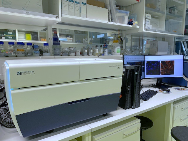
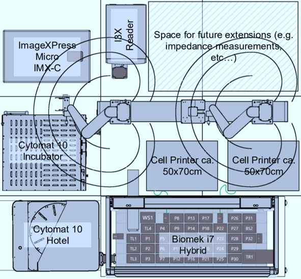
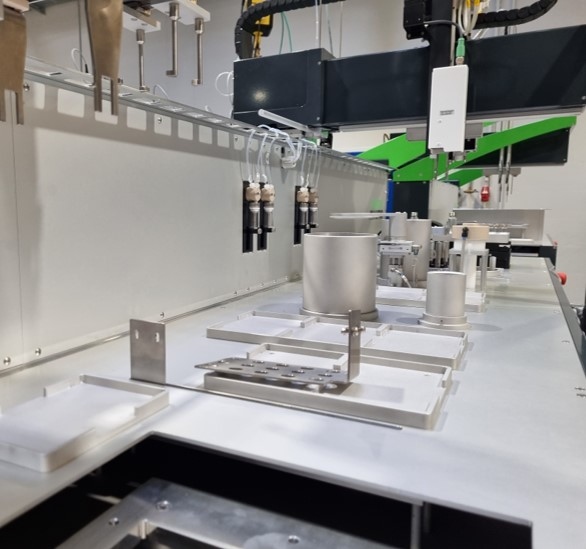
ImageXPress HT-AI: Fully automated confocal (spinning disc) HT-screening and 3D tissue imaging pipeline, containing an ImageXpress HT-AI boxed microscope. The ImageXpress® Confocal HT.ai High-Content Imaging System employs a seven-channel laser light source along with eight imaging channels, facilitating highly multiplexed assays while ensuring rapid throughput through reduced exposure times. Water immersion objectives enhance image clarity and reduce distortions, enabling the deeper penetration into dense samples. The powerful combination of MetaXpress® software and IN Carta® software simplifies workflows for advanced phenotypic classification and 3D image analysis with machine learning capabilities and an intuitive user interface. The ImageXpress HT-AI is connected to a fully automated incubation (Thermofisher Cytomat), pipetting (Opentron, Eppendorf EP5075), spectrophotometry (Sprectramax ID3), automated FACS (Guava easyCyte HT) pipeline handled by a Precision Flex 400 robotic arm system (Brooks). The system is also connectect to an HT-DLP 3D-printer (BioNOVAX) and a contactless rheometer (Rheolution) for generating 3D tissues and biomaterials testing. The pipeline is part of the the self driving lab Bio.Mat, which is embedded in the Auto.MAP alliance. Auto.MAP is the accelerated materials platform at KIT.
Contact: Ute Schepers
Leica MICA Microhub
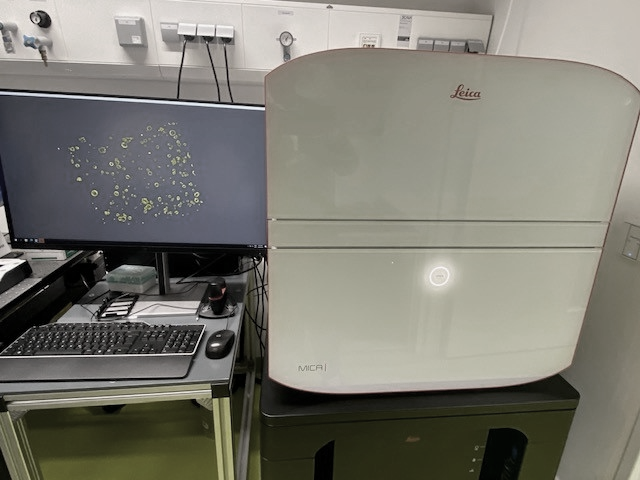
The Mica Microhub is a highly automated boxed system that combines widefield and confocal imaging within a sample-protecting, incubating environment. It significantly expediting fluorescence imaging workflows and yielding meaningful scientific results at a faster pace and allows for large screening experiments for cells and 3D tissues. The Microhub feature allows for the simultaneous capture of all four labels representing different structures in a single acquisition, whether for widefield or confocal imaging, eliminating the need to move the sample. This effectively addresses the spatiotemporal mismatch between labels of moving objects encountered during sequential acquisition. Furthermore, all these functionalities are driven by the patented FluoSync technology, offering a swift and gentle approach to multicolor fluorescence imaging.
Contact: Véronique Orian-Rousseau
Leica Thunder Imager HT
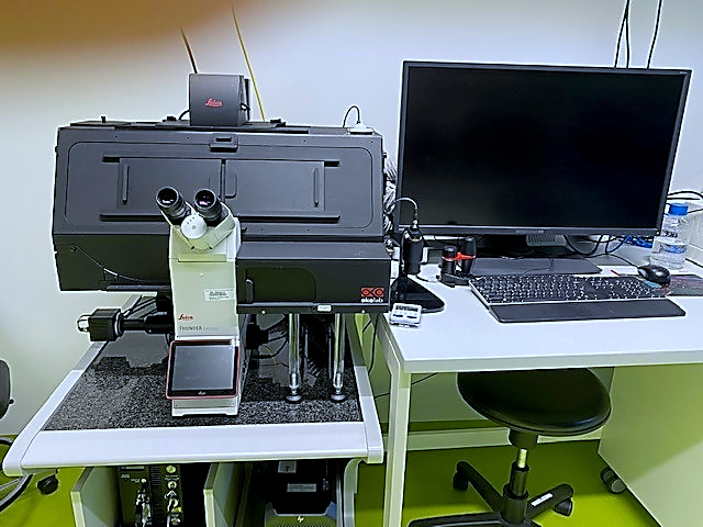
THUNDER Imaging Systems empower users to achieve precise visualization of intricate details, even within deeply embedded samples, in real-time, devoid of any out-of-focus blurring. THUNDER delivers high-speed, multi-color imaging of samples of varying thickness, providing enhanced temporal resolution right from the initial attempt. Obtaining sharp images of three-dimensional specimens becomes as effortless as operating your preferred camera-based fluorescence microscope. TheThunder Software enables clear view of details even deep within the specimen due to Computational Clearing, directly in live preview.
Contact: Anna Popova, Pavel Levkin