
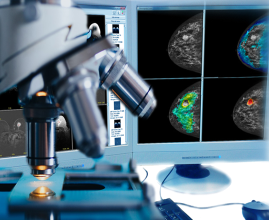
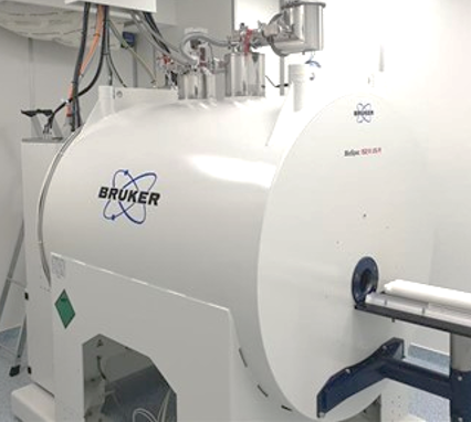
Medical imaging and image processing play a central role in medical diagnostics and therapy. Multimodal data from a wide variety of imaging methods, large data volumes due to increasingly higher image resolution, and three-dimensional images are just some of the challenges faced by scientists in differential diagnostics. With the Tissue Imaging Center we have established a research platform to accelerate research and development of a broad spectrum of screening and imaging methods. In collaborative research projects we are developing novel and cutting-edge technologies on tissue imaging and image data processing ranging from high resolution and high throughput microscopical imaging to correlative imaging, NMR, X-ray, sophisticated MRI and ultrasound technologies. For cell biology the Tissue Imaging Center is dedicated to provide solutions for imaging of all kinds of biological specimens from single
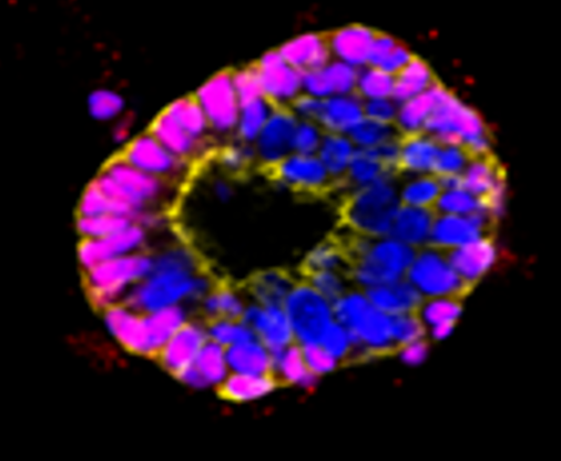
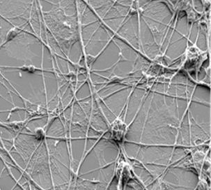
organelles in high resolution up to 3D tissues and whole organs with exceptional speed and precision. Whether you're exploring the intricacies of organoid biology, conducting large-scale drug screenings, or seeking to understand complex tissue interactions, our state-of-the-art technology and expertise are here to support your research endeavors in collaborative research projects. With our fully automated High- Throughput Screening capabilities, we have established efficient and streamlined imaging processes, allowing for the rapid analysis of a large number of samples. Our automated microscopy systems are equipped to handle various types of organoids and tissue specimens, enabling high-resolution confocal and digital light sheet imaging across multiple experimental conditions. From experimental design and sample preparation to data acquisition and analysis, we provide comprehensive support tailored to your specific needs. At the Tissue Imaging Center, we are dedicated to empowering researchers to push the boundaries of scientific discovery. We look forward to collaborating with you and contributing to the advancement of your research in cell to tissue imaging and organoid biology.
Tissue Imaging Center
Our Tissue Imaging Center is a research platform, which provides advanced imaging solutions for analyzing tissues and cells with high throughput capabilities and high precision.
Research Platform for Technology Development
Imaging Equipment
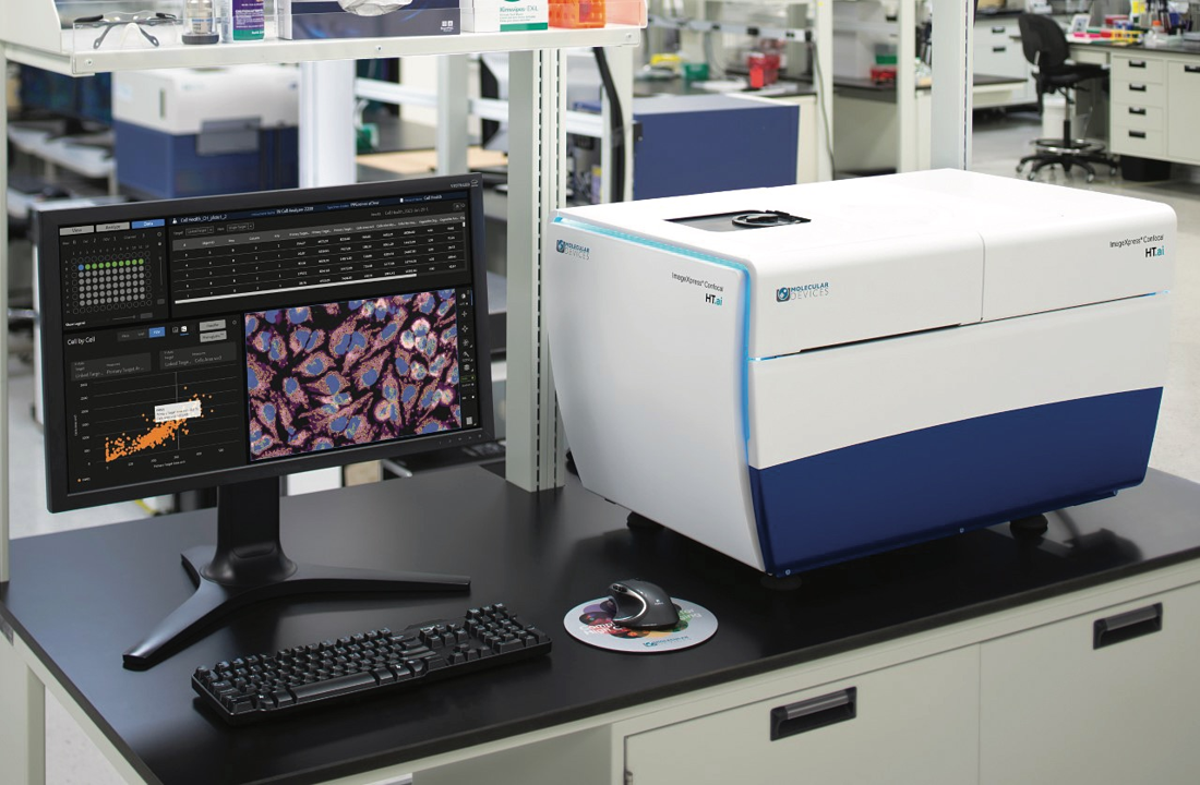
- ImageXPress HT-AI: Fully automated confocal (spinning disc) HT-screening and 3D tissue imaging pipeline, containing an ImageXpress HT-AI boxed microscope. The ImageXpress® Confocal HT.ai High-Content Imaging System employs a seven-channel laser light source along with eight imaging channels, facilitating highly multiplexed assays while ensuring rapid throughput through reduced exposure times. Water immersion objectives enhance image clarity and reduce distortions, enabling the deeper penetration into dense samples. The powerful combination of MetaXpress® software and IN Carta® software simplifies workflows for advanced phenotypic classification and 3D image analysis with machine learning capabilities and an intuitive user interface. The ImageXpress HT-AI is connected to a fully automated incubation (Thermofisher Cytomat), pipetting (Opentron, Eppendorf EP5075), spectrophotometry (Sprectramax ID3), automated FACS (Guava easyCyte HT) pipeline handled by a Precision Flex 400 robotic arm system (Brooks). The system is also connectect to an HT-DLP 3D-Printer (BioNOVAX) and a contactless rheometer (Rheolution) for generating 3D tissues and biomaterials testing.
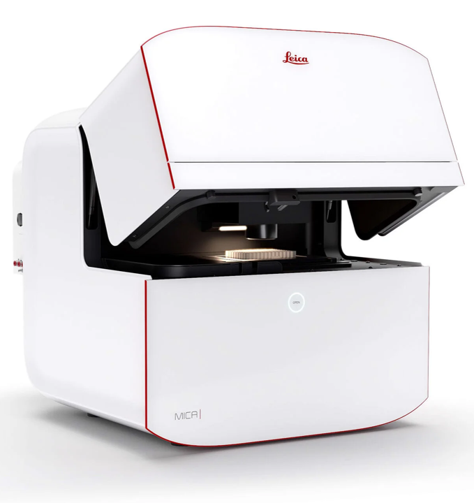
- Leica Mica Microhub: The Mica Microhub is a highly automated boxed system that combines widefield and confocal imaging within a sample-protecting, incubating environment. It significantly expediting fluorescence imaging workflows and yielding meaningful scientific results at a faster pace. The Microhub feature allows for the simultaneous capture of all four labels representing different structures in a single acquisition, whether for widefield or confocal imaging, eliminating the need to move the sample. This effectively addresses the spatiotemporal mismatch between labels of moving objects encountered during sequential acquisition. Furthermore, all these functionalities are driven by the patented FluoSync technology, offering a swift and gentle approach to multicolor fluorescence imaging.
- Leica Thunder Imager HT: THUNDER Imaging Systems empower users to achieve precise visualization of intricate details, even within deeply embedded samples, in real-time, devoid of any out-of-focus blurring. THUNDER delivers high-speed, multi-color imaging of samples of varying thickness, providing enhanced temporal resolution right from the initial attempt. Obtaining sharp images of three-dimensional specimens becomes as effortless as operating your preferred camera-based fluorescence microscope. TheThunder Software enables clear view of details even deep within the specimen due to Computational Clearing, directly in live preview.
- Leica Stellaris Digital Light Sheet (DLS) confocal system: STELLARIS 5 Digital LightSheet (DLS) combine a confocal system and a light sheet microscope in one, providing a unique and versatile tool for high resolution imaging of 3D tissues and organoids. The distinctive vertical design of DLS, made possible by Leica Microsystems’ proprietary TwinFlect mirrors, enables the integration of confocal and light sheet imaging within the same system. This allows you to easily adapt your microscopy approach to suit the specific requirements of your experiments. DLS enhances the flexibility of your research by accommodating various sample types, including model organisms, organoids, or cleared tissue, and by leveraging the full excitation spectrum. This adaptability is facilitated by the STELLARIS White Light Laser and the capability to conduct multi-position light sheet experiments using standard glass bottom plates. These features enable you to explore your research questions with new and improved methodologies.
Leica Stellaris 5 TauSense system: The Stellaris 5 confocal system is equippedwith the TauSense unit to allow for lifetime-based information for enhanced multiplexing and functional imaging
- Zeiss LSM 800
- Zeiss Cell observer:
- Keyence microscopes